In recent years, volumetric CAD packages have emerged as the tool of choice for medical modeling. Geomagic Freeform, one such volumetric CAD package, is well suited to give designers speed and design freedom with complex, organically shaped objects such as those found in the human body.
By Kevin Atkins
3D Systems Geomagic
When a defect, deformity, or injury alters cranial bones, surgeons need a unique blend of engineering and artistry to return the human face to its former aesthetics. Advances in volumetric CAD technology are allowing traditional CAD manufacturing principles to meet complex medical modeling needs that formerly couldn’t be handled digitally — like reshaping the human skull. These advances let biomedical engineers digitally sculpt a custom cranial implant or surgical guide with the best fit and function for each patient.
A new breed of rapid-product-development firms provides doctors and patients with such surgical guides and custom implants faster and more cost-effectively, thanks to advances in rapid manufacturing techniques and biocompatible materials.
Despite readily available digital medical imaging files such as CT scans and MRIs, patient-specific implants and surgical guides are still primarily made by hand by small labs. Organizations usually can’t support volume demand for digital fabrication devices to justify their cost. Labs cut, grind and manually shape surgical guides and implants that must be iteratively fitted to the patient, despite the fact that there are precise digital files that represent the perfect template for the desired human form. In the case of a cranial implant, time is often of the essence, minimizing the patient’s risk of further injury or infection.
Medical design specialist firms and small biomedical device makers have made it possible for specialty domains, such as cranial implants and surgical guides, to access technologies that were previously out of the reach of small in-hospital labs or even large implant design and manufacturing firms.
These specialists can provide devices on an as-needed basis, supporting the limited runs required for custom needs, without requiring the overhead of buying and maintaining additive manufacturing 3D printers or rapid prototyping (RP) systems in-house. Custom surgical guides and implants can cut the cost of healthcare services downstream by improving surgical predictability and reducing operating room time for the maximum number of patients. And rising health care costs and the fact that insurance companies limit coverage means that reducing overhead costs is paramount.
Volumetric CAD: a Better Way
The broad range of clinical cases requiring craniofacial implants and prosthetic reconstruction requires software that provides a high degree of design flexibility and is intuitive to use, says Dominic Eggbeer, research officer and head of medical applications at the Centre for Applied Reconstructive Technologies in Surgery (CARTIS), in Swansea, UK, (cartis.org) and affiliated with Morriston Hospital. CARTIS has worked for years with expert research groups around the world to discover innovative ways to rebuild damaged facial anatomy.
In recent years, volumetric CAD packages have emerged as the tool of choice for medical modeling. Traditional CAD, designed for engineers, creates geometric, multipart products and often takes too long to render organic shapes to be practical. But volumetric or sculptural CAD packages provide medical specialists, who are both artist and anatomy experts, with a great bridge to the digital world. The typical “carve here, smooth there” work that designers formerly did by hand to ensure the perfect fit can be done digitally, directly on the 3D model, more quickly and precisely. Designers can harness the mathematical precision and speed of digital engineering tools to design shapes that are organic, sculptural and asymmetrical.
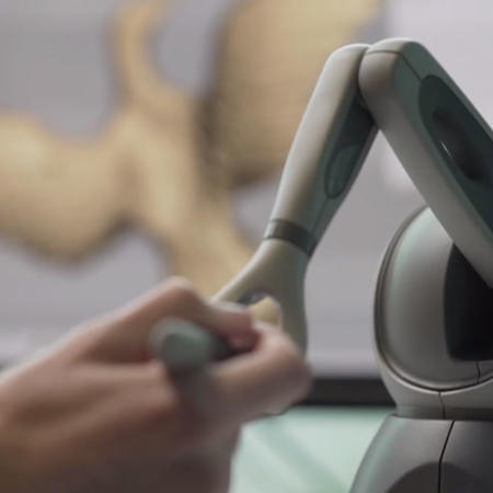
The Geomagic® Freeform® design system is one example of a volumetric CAD package and is based on voxels, or 3D pixels. Voxels stick together to create a solid mass and can be sculpted just like clay or wax. As a result, the software is well suited to give designers speed and design freedom with complex, organically shaped objects such as those found in the human body.
Brain Surgery for Infants
In one example of the software’s use, several times each month, as many as 30 surgeons and medical experts gather in Hershey, Pa., at Penn State’s College of Medicine (pennstatehershey.org) for Surgical Innovation Group (SIG) meetings. The purpose of the meetings is to generate a vibrant discussion about challenges in day-to-day surgical and medical care. The discussions are a process that generates ideas, projects and eventually devices that have been vetted for their value and likelihood to reach the marketplace and benefit patients. Participating in the discussions is Thermoplastic Products Corporation (TPC) of Hummelstown, Pa., whose principal is Barry Fell. When the topic of craniosynostosis, or grossly misshapen infant heads, arose recently, Fell had an elegant answer that typifies the way volumetric CAD is advancing medical treatment.
In craniosynostosis, the baby’s cranial bones are prematurely fused and, fueled by the underlying brain?growth, the? skull becomes misshapen. The premature fusion of the skull bones can also lead to increased intracranial pressure on the brain, swelling of the optic disc and vision loss, and potential long-term developmental problems. The only treatment option is surgery. The skull bones are cut to release the fused structure, and then they are reshaped and placed back on the patient much like large puzzle pieces. The surgeon accounts for the increased space needed for the brain to grow by careful bone placement and fixation.
In the past, surgeons would plan craniosynostosis surgeries by reviewing infants’ CT scans and formulating a generic game plan. However this approach would offer only an approximate guide to the infant’s unique anatomy, and more importantly, it would defer determining much of the surgical plan until the patient’s cranium was open and exposed during surgery.
Since most craniosynostosis patients receive a CT scan of the head preoperatively, Fell decided to leverage volumetric CAD and FEA technology to help both surgeon and patient.
A surgeon working on a craniosynostosis case delivered a conventional CT scan from an infant patient to TPC, which converted it into an STL format. Engineers imported the STL file into Freeform and began by cleaning up “noise,” or artifacts, from the original CT scan to more clearly identify the cranial fissure lines. Engineers then used Freeform’s AutoSurfacer to create a surface mesh — a needed step to ensure the evolving design would accurately capture the varying thicknesses of the skull.
In past attempts, Fell needed between two to four hours to create a mesh file of a curved, organic shape with varying thickness in a traditional CAD package. Other software could not surface the parts because of their complexity. Therefore, the attempt would go on for a long time and then fail.
But by using Freeform, Fell’s team cleaned up the STL file, performed the autosurfacing, and completed a final design in about 30 minutes. Freeform’s AutoSurfacer feature worked well on even the most unusual shapes that TPC needed.
For the corresponding surgical guide, TPC’s team took the Freeform file and used it as their source for an FEA projection of the baby’s head shape if it was allowed to grow several months without surgery. From FEA predictions of how the bones would be shaped in the future, surgeons had a highly accurate, digital model from which to plan specific surgical bone cuts to allow the optimal reassembled cranium.
Working together over a computer, the surgeon and TPC engineer then “fixed” the skull by performing surgical cuts on the bone — using Freeform — and placed the bone back into the anticipated post-surgical position. TPC then adjusted the design file, repeated the FEA, and confirmed that the new shape would have uniform stress predictions throughout the skull so that it would maintain the desired shape as the baby’s brain grows.
Once engineers determined the final skull shape, Freeform helped TPC design a surgical template for the surgeon to guide the bone cuts for the ideal skull shape. The surgeon actually lays a patient-specific template on top of the bone to guide the cuts.
After surgeon review and approval of the surgical guide, TPC confirmed final placement of the patient-specific template on the skull model and predicted the future shape. The team then used a rapid-prototyping machine to build the disposable template out of an FDA-approved biocompatible resin for the actual surgery.
For Trauma Patients
In another example, MedCAD, a biomedical-device manufacturer in Dallas, provides custom cranial implants for infants and adults. It offers surgical guides and a new line of cranial implants called AccuShape, which address conditions similar to those facing Rep. Gabrielle Giffords, the victim of a gunshot wound to the head. In this patient’s case, the brain’s swelling during the stroke required removing a section of the skull and replacing it with an implant.